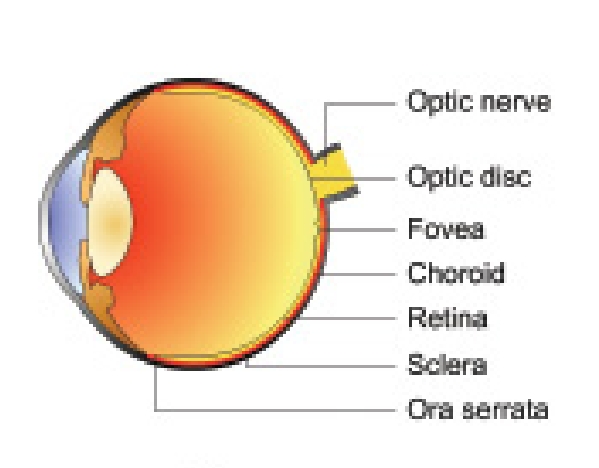
Retina, the innermost tunic of the eyeball, is a thin, delicate and transparent membrane. It is the most highly-developed tissue of the eye. It appears purplish-red due to the visual purple of the rods and underlying vascular choroid.
Gross anatomy
Retina extends from the optic disc to the ora serrata. Grossly it is divided into two distinct regions:
posterior pole and peripheral retina separated by the so called retinal equator.
Retinal equator is an imaginary line which is considered to lie in line with the exit of the four vena verticose.
Posterior pole refers to the area of the retina posterior to the retinal equator.
The posterior pole of the retina includes two distinct areas: the optic disc and macula lutea .
Posterior pole of the retina is best examined by slit-lamp indirect biomicroscopy using +78D and +90D lens and direct ophthalmoscopy.
Optic disc.
It is a pink coloured, well-defined circular area of 1.5-mm diameter. At the optic disc all the retinal layers terminate except the nerve fibres, which pass through the lamina cribrosa to run into the optic nerve.
A depression seen in the disc is called the physiological cup. The central retinal artery and vein emerge through the centre of this cup.
Macula lutea.
It is also called the yellow spot. It is
comparatively deeper red than the surrounding fundus and is situated at the posterior pole temporal to the optic disc. It is about 5.5 mm in diameter. Fovea centralis is the central depressed part of the macula.
It is about 1.5 mm in diameter and is the most sensitive part of the retina. In its centre is a shining pit called foveola (0.35-mm diameter) which is situated about 2 disc diameters (3 mm) away from the temporal margin of the disc and about 1 mm below the horizontal meridian.
An area about 0.8 mm in diameter (including foveola and some surrounding area) does not contain any retinal capillaries and is called foveal avascular zone (FAZ). Surrounding the fovea are the parafoveal and perifoveal areas.
Peripheral retina refers to the area bounded posteriorly by the retinal equator and anteriorly by the ora serrata. Peripheral retina is best examined with indirect ophthalmoscopy and by the use of Goldman three mirror contact lens.
Ora serrata. It is the serrated peripheral margin where the retina ends. Here the retina is firmly attached both to the vitreous and the choroid. The pars plana extends anteriorly from the ora serrata.
Microscopic structure
Retina consists of 3 types of cells and their synapses arranged (from without inward) in the following ten layers
- Pigment epithelium
- Layer of rods and cones
- External limiting membrane
- Outer nuclear layer
- Outer plexiform layer
- Inner nuclear layer
- Inner plexiform layer
- Ganglion cell layer
- Nerve fibre layer
- Internal limiting membrane
1. Pigment epithelium.
It is the outermost layer of retina. It consists of a single layer of cells containing pigment. It is firmly adherent to the underlying basal lamina (Bruch’s membrane) of the choroid.
2. Layer of rods and cones.
Rods and cones are the end organs of vision and are also known as photoreceptors. Layer of rods and cones contains only the outer segments of photoreceptor cells arranged in a palisade manner.
There are about 120 millions rods and 6.5 millions cones. Rods contain a photosensitive substance visual purple (rhodopsin) and subserve the peripheral vision and vision of low illumination (scotopic vision).
Cones also contain a photosensitive substance and are primarily responsible for highly discriminatory central vision (photopic vision) and colour vision.
3. External limiting membrane.
It is a fenesterated membrane, through which pass processes of the rods and cones.
4. Outer nuclear layer.
It consists of nuclei of the rods and cones.
5. Outer plexiform layer.
It consists of connections of rod spherules and cone pedicles with the dendrites of bipolar cells and horizontal cells.
6. Inner nuclear layer.
It mainly consists of cell bodies of bipolar cells. It also contains cell bodies of horizontal amacrine and Muller’s cells and capillaries of central artery of retina. The bipolar cells constitute the first order neurons.
7. Inner plexiform layer.
It essentially consists of connections between the axons of bipolar cells dendrites of the ganglion cells, and processes of amacrine cells.
8. Ganglion cell layer.
It mainly contains the cell bodies of ganglion cells (the second order neurons of visual 7pathway). There are two types of ganglion cells. The midget ganglion cells are present in the macular region and the dendrite of each such cell synapses with the axon of single bipolar cell.
Polysynaptic ganglion cells lie predominantly in peripheral retina and each such cell may synapse with upto a hundred bipolar cells.
9. Nerve fibre layer (stratum opticum)
It consists of axons of the ganglion cells, which pass through the lamina cribrosa to form the optic nerve. For distribution and arrangement of retinal nerve fibres
10. Internal limiting membrane.
It is the innermost layer and separates the retina from vitreous. It is formed by the union of terminal expansions of the Muller’s fibres, and is essentially a basement membrane.


